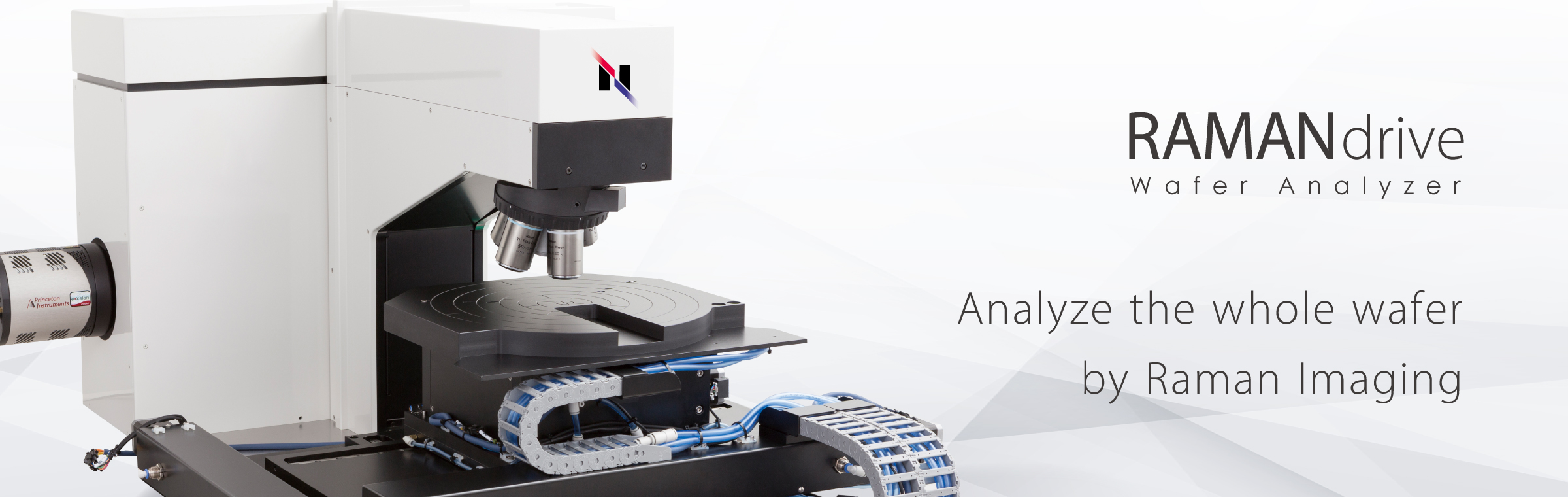
RAMANdrive is the specialized Raman microscope for wafer analysis equipped with our dedicated 300 mm stage. RAMANdrive gives you an ultra-fast, highest resolution analysis of the whole wafer with unique stability and accuracy
Stage Navigation System
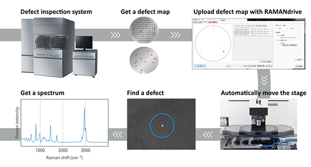
The Nanophoton Stage Navigation System accepts data from your inspection system and use it to move the wafer to all indicated positions for a detailed analysis. The dedicated stage moves the wafer safely and with high accuracy to all areas of interest.
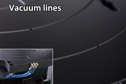
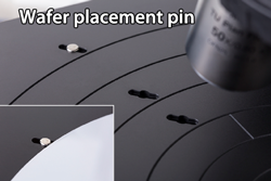
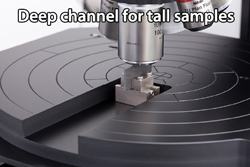
Accurate and stable 300 mm wafer stage
Large wafers of up to 300 mm can be placed on the stage. Vacuum lines are connected to small holes in channels, which allows a applied vacuum to flatten and hold the wafer on the stage. Our extended wafer stage makes it possible for the laser to access the entire exposed wafer surface. Even a tall sample also can be analyzed by placing it in the deep channel part of the stage.
Upload your areas of interest with the Stage Navigation System
The Stage Navigation System is an essential part of the RAMANdrive software, designed to save time and improve the efficiency. Simply upload your data from your regular inspection tool and RAMANdrive identifies your areas of interest and moves the wafer automatically to the requested positions for a detailed analysis. This technology works even with other samples if requested
High Performance Raman Imaging
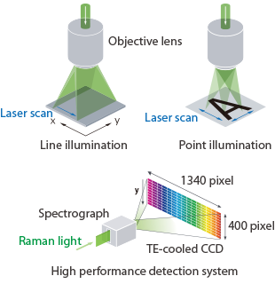
Nanophoton developed a dedicated technology providing highest speed by using a unique laser line illumination system. A special illumination and detection system gets 400 spectra with only one laser shot. By moving the laser line across your sample without moving the sample, several hundred thousands of data are collected within a few minutes to complete a whole Raman image of your area of interest. The Nanophoton technology provides highest speed without limitation in resolution and accuracy.
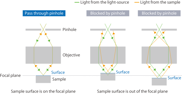
Nanophoton developed a dedicated confocal optical system providing highest spatial and spectral resolution in 3 dimensions. The highest spatial resolution enhances the sensitivity to detect particles of less than 100 nm. Highest spectral resolution combined with the necessary positioning accuracy gives you tremendous data even for stress or polytype analysis.
Highest spatial resolution
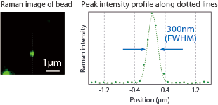
| Sample | Fluorescent beads(Φ200 nm) |
| Excitation wavelength | 532 nm |
| Obj. lens | 100x, 0.90 NA |
Spectral resolution and peak positioning accuracy
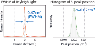
| Excitation wavelength | 785 nm (L) | 532 nm (R) |
| Grating | 1200 (L) | 2400 (R) |
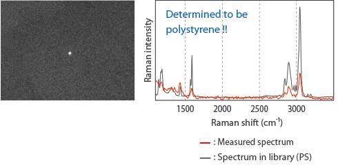
Raman analysis of 100 nm particle
Our highest spatial resolution enhances the sensitivity to detect tiny particles. High-quality dark-field microscopy can easily identify particles of less than 100 nm. The laser beam will then be accurately focused on it using galvanometer mirrors, producing a spectrum with a high signal-to-noise ratio that an identification can be done by a library search
Visualize stress distribution by 3D Raman Imaging
Our confocal optics allows depth profiling of transparent samples such as SiC and GaN. Here we demonstrate the cross-sectional Raman imaging of stress distribution in a SiC wafer. It easily shows that stress is reduced by the polishing process.
Photoluminescence (PL) imaging is widely used to observe the distribution of defects, impurities and GaN itself. 325 nm UV laser option is used to surpass the band gap and detect PL spectra of InGaN dots with different sizes and composition. A UV laser is also useful to measure Raman spectra at the outermost surface of samples because of its short penetration depth.
Application note
→SiC(シリコンカーバイド)の応力評価
Application note
→紫外励起PLイメージングとラマンイメージング
Specification
Performance
| SPATIAL RESOLUTION(X / Y / Z) | 350nm / 500nm / 1000nm(@532nm、100x 0.90NA) |
|---|---|
| SPECTRAL REGION | 100cm-1~ (option:50 cm-1~) |
| SPECTRAL RESOLUTION(FWHM) | 1.2cm-1 (@785nm、1200gr/mm) |
| PIXEL RESOLUTION(SPECTRUM) | 0.4cm-1/pixel (@785nm、1200gr/mm) |
| PEAK POSITION ACCURACY | 0.1cm-1 (@532nm、2400gr/mm) |
Specification
| LASER WAVELENGTH | 325nm, 355nm, 488nm, 532nm, 671nm, 785nm, others |
|---|---|
| ILLUMINATION TYPES | Point illumination / Line illumination |
| FOCAL LENGTH OF SPECTROMETER | 550mm |
| GRATING | 150, 300, 600, 1200, 1800, 2400gr/mm(Max. 3 gratings, selectable) |
| DETECTOR | TE-cooled CCD 1340 × 400 pixel format |
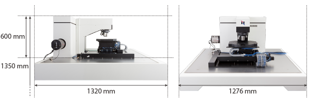
Size and Weight
| SIZE(W×H×D) | W1276 × D1320mm × H1350 (Including anti-vibration table) |
|---|---|
| WEIGHT | 390kg (Including anti-vibration table) |