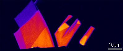
■: Single-layer graphene
■: Double-layer graphene
■: Triple-layer graphene
■: Quadruple-layer graphene
| Excitation wavelength | 532 nm |
| Obj. lens | 100x (NA=0.90) |
| Number of spectra | 67,600 (400×169) |
| Measurement time | 5 min 30 sec |
Raman image of more than 60,000 spectra can be obtained in just 5 minutes
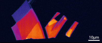
The image above shows the Raman imaging of graphene on a heat oxidized silicon substrate. It takes only 5 minutes to identify and visualize the distribution of single-layer graphene which is one carbon atom sheet and double-layer, triple-layer and quadruple-layer graphene respectively.
■About the sample used for measurement
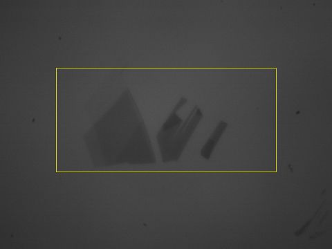
This sample is prepared by cleaving graphite with adhesive tape then transcribing it into silicon substrate. In this way, all graphene are formed on silicon substrate at random, and if the substrate has a certain amount of SiO2 film on it, they can be detected by optical microscopy. However, it is difficult to distinguish exactly how many layers each graphene consists of (shown in the right image).
*This sample is provided by Dr. Daiju Tsuya of National Institute for Materials Science.
References
K.S. Novoselov et al., Science 306, 666 (2004).
K.S. Novoselov et al., Proc. Natl. Acad. Sci. U.S.A.102, 10451 (2005).

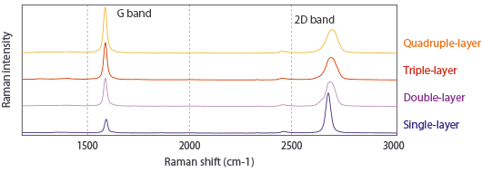
Identification of number of graphene layer by G/2D ratio
The number of graphene layers can be identified by investigating the intensity ratio of G-band and 2D-band in the Raman spectrum of graphene. In single-layer graphene, a very sharp strong peak can be seen in 2D-band. Moreover, peak intensity in G-band becomes higher as the number of layers increases.


G/2D is about 0.3 in single-layer and increases linearly until quintuple layers and saturated in more than sextuple layers. In single-layer graphene, the peak position of 2D-band is at about 2678.8 ± 1.0 cm-1, but when it becomes more than double layers, the peak position of 2D-band shifts to higher wavenumber and the FWHM becomes broad. Moreover, when the graphene becomes more than double layers, 2D-band is composed of two or more subpeaks, while 2D-band of single-layer graphene can be fitted with Single Lorentzian. That is why the wave forms on the low wavenumber side of 2D-band in double-layer graphene have a little distortion of shape. As described above, a lot of useful information about graphene structure is included in the Raman spectrum of graphene.
References
D. Graf et al., Nano Letters. 7, 238 (2007).