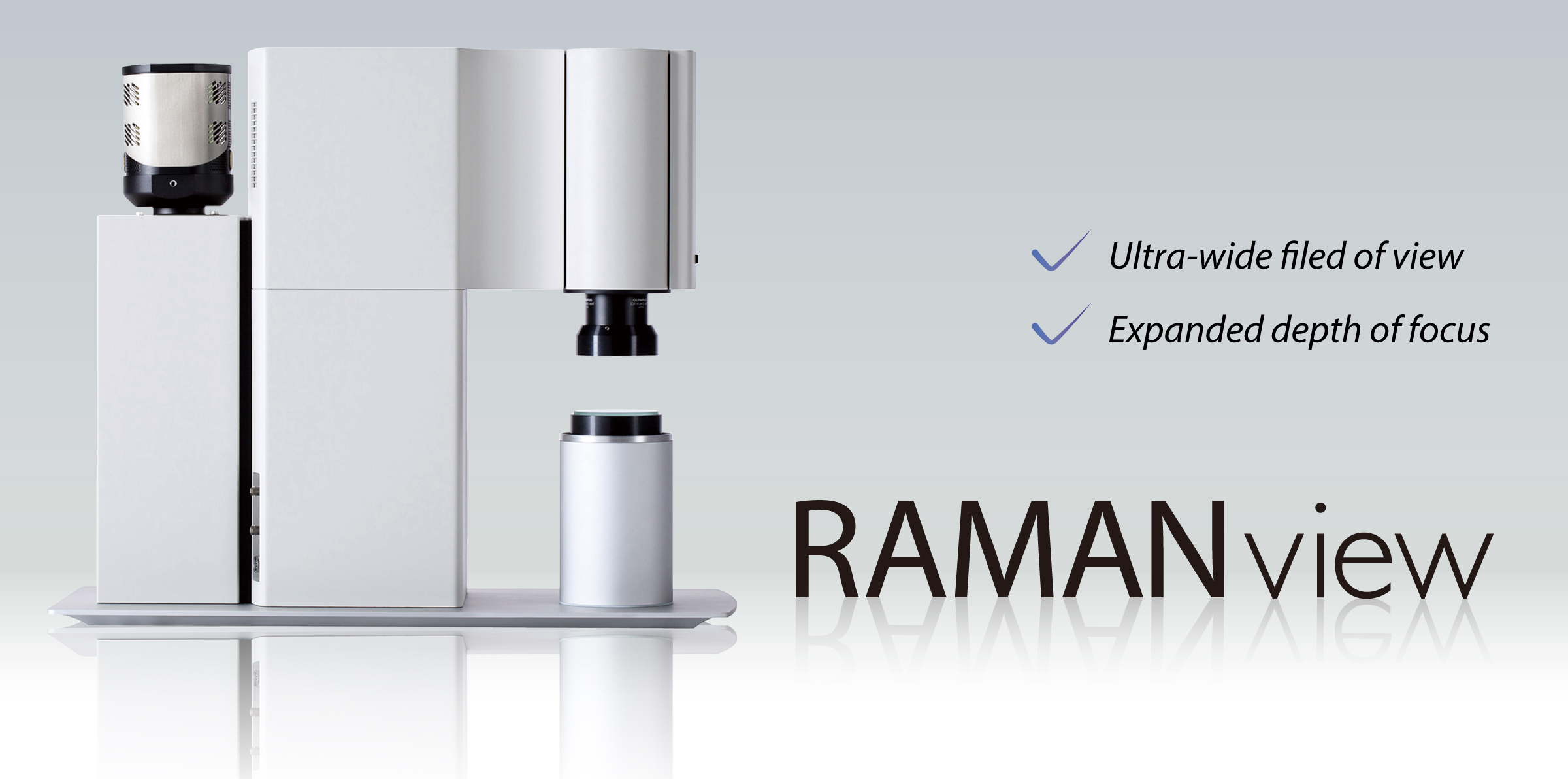
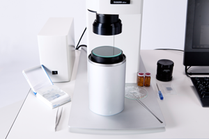
What is RAMANview?
Substantially extend your view with a unique Raman imaging system. RAMANview provides you a whole new dimension for easy to use operation and ultra-fast screening!
The one and only performance
“Ultra-wide field-of-view”, “Extended depth of focus”, “High spatial resolution”, “Ultra-long working distance”.
These 4 values inside the minimum body enable diverse Raman spectroscopic analysis.
More than 800 mm2 is analyzed without moving the sample.
RAMANview has a dedicated optical system, which substantially expand the field-of-view. Even large samples – bigger than 800 mm2 – can be analyzed without moving the sample. Just select with the mouse of your computer the area of interest and the ultra-fast laser scanning system will do the requested analysis. Regular mapping systems take a long time; have the risk of moving the sample out of area or ? working with solutions ? even slopping it. RAMANview saves your time and provides a secure analysis of any area of interest.
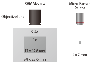
Example of field size of wide-field Raman imaging
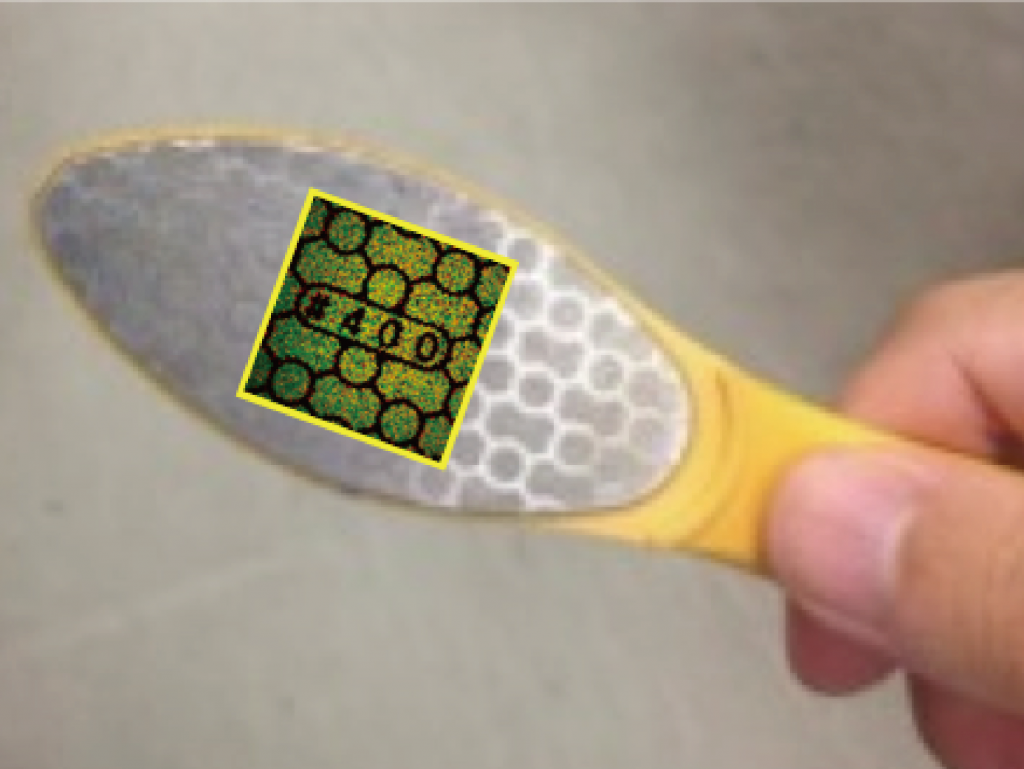
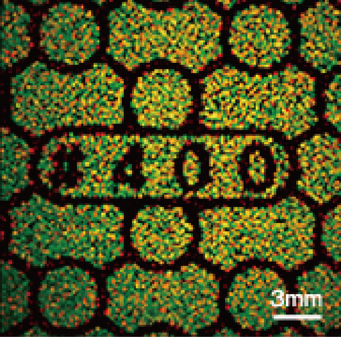
Left: The sample (diamond file) and measurement area
Right: Wide-field Raman image
Be free from time-consuming focus corrections by extended focal depth
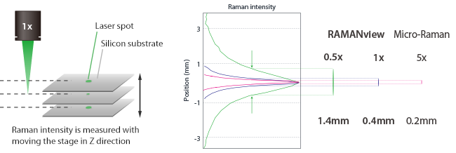
The dedicated optical system of RAMANview provides an extended depth of focus of up to about 1.4 mm (with the 0.5x objective) for samples even with a height difference of 1 or 2 mm without annoying and misty out of focus areas. This unique depth of focus enables easy-of-use sampling without sectioning or cutting the sample.
The relationship between magnification of objective lens and depth of focus
Tiny, micro scale components will be detected with RAMANview
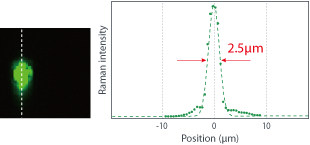
The dedicated optical system creates a high precision spot size, which facilitates a spatial resolution of up to 2.5 μm (see right figure) with the 2x objective lens. RAMANview also stands for very fast Raman imaging in a micro scale range.
Spatial resolution assessment of RAMANview
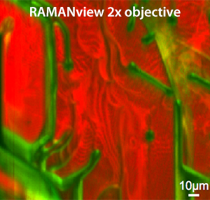
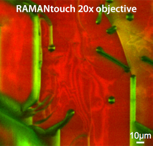
■4H-SiC ■6H-SiC
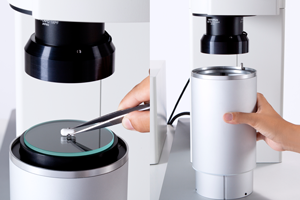
Ultra-long working distance extends the measurement scene.
RAMANview has a working distance of more than 70 mm (with the 0.5x objective). It offers an easy-to-use sampling (safe distance between lens and sample) and allows measurements even in deep containers, vessels or in extensive sampling systems. Even measurements through windows or transparent films etc. are not an issue. RAMANview gives you the space you need for your sampling!
Measurement of a sample in a H 45mm vial
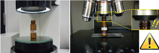
Left: By RAMANview.
Right: By conventional Raman microscope.
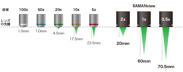
The relationship between magnification of objective lens and working distance
Whole new applications
RAMANview, the wide-field Raman microscope has changed the common knowledge of space scaling of Raman imaging. A brand new world of application has been broadened.
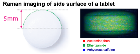
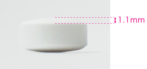
High speed Raman imaging analysis of a tablet
RAMANview is a perfect tool for Raman imaging of tablets even with a 1 mm difference in height. Within only 14 minutes, an area of about 100 mm2 was analyzed with the 1x objective. A greater difference can be done with the 0.5x objective. Nanophoton’s laser line scanning technology creates this unsurpassed speed with excellent high precision.
Raman imaging of the whole surface of a tablet
Measurement condition (left)
| Excitation wavelength | 532 nm |
| Obj. lens | 1x |
| Pixels | 202 x 202 pixel |
| Measurement time | 14 min |
Measurement condition (right)
| Excitation wavelength | 532 nm |
| Obj. lens | 0.5x |
| Pixels | 99 x 192 pixel |
| Measurement time | 8 min |
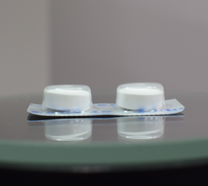
Raman imaging of a tablet inside a blister pack
Even the imaging and analysis of a tablet inside a blister pack is possible by RAMANview. The fixation of tablet inside the pack cannot be flattened by shaving. Fixation is also impossible, so by moving the stage the tablet will swing inside the blister pack and a Raman analysis would not be possible. Only the unique technology of RAMANview can perform fast imaging under such conditions.
| Excitation wavelength | 532 nm |
| Obj. lens | 1x |
| Pixels | 202 x 202 pixel |
| Measurement time | 14 min |
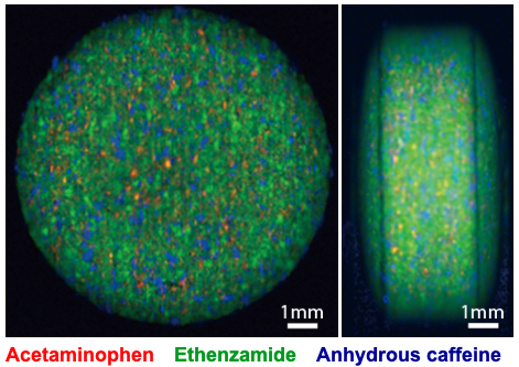
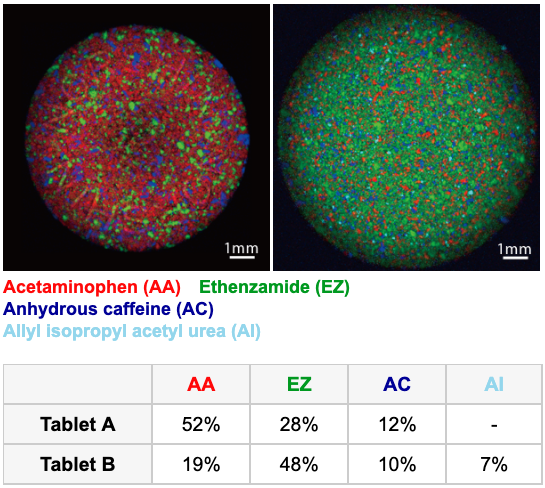
Highly precise Raman imaging analysis of tablet
High-definition Raman imaging, which is realized by point illumination with small scanning steps, is recommended for observing distribution of components precisely. The data shown on the right are the high-definition Raman images of 2 different types of antipyretic tablets. Fast laser beam scanning provides high imaging speed. The component distribution is clearly observed by the high spatial resolution over a wide range. Going even beyond, for a small amount of components, like the API’s in tablets, an additional particle analysis is available. The software identify the particles one by one and a statistical analysis of diameter, area and volume is provided.
High-definition Raman images of two different tablets
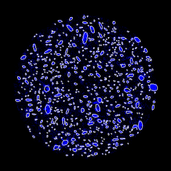
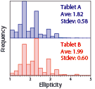
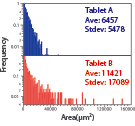
Particle size analysis of caffeine during formulation (comparison between pharmaceutical A and B)

Ellipticity histogram
Area histogram
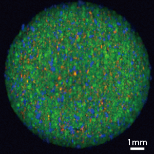
Detect 1% API among the pharmaceutical formulation content
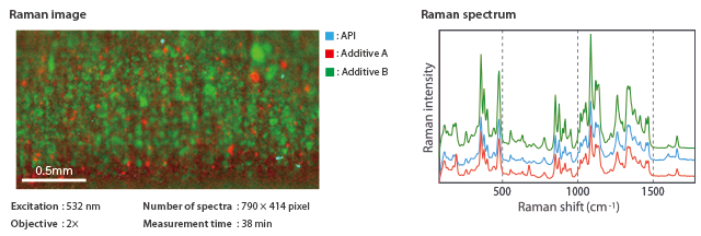
This shows the component distribution of a tablet. RAMANview’s 2x objective lens is able to detect the slight amount of APIs (0.5mg) during solid dosage form by means of both a high spatial resolution and wide-area scanning.
Analysis of powder inside a plastic bag
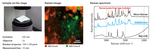
The deep depth of focus is a great tool for bulky samples, however, even an analysis inside thin plastic bags are greatly performed with RAMANview. Raman peaks from thin film, which can be a noise for micro-Raman system, are not included in a spectrum obtained by RAMANview.
Analysis of crystallinity distribution of resin molded product

The FWHM of polyethylene terephthalate’s peak at around 1720cm-1 changes due to the degree of crystallinity. The image above visualizes the difference in the degree of crystallinity from the mouth to the body part of PET bottle.
Observation of the composition distribution of Inada granite’s surface
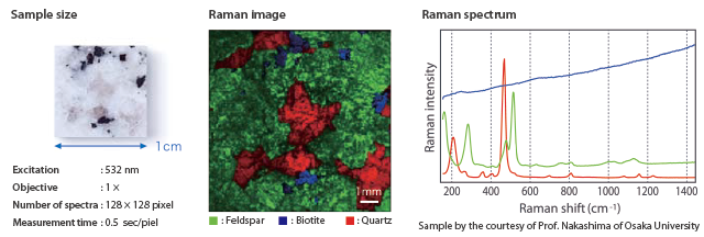
The red, blue and green parts are known as quartz, biotite and feldspar respectively from the Raman spectrum. Deterioration and erosion of the rock can be captured with a centimeter-order field-of-view by RAMANview.
※This sample is provided by Professor Satoru Nakajima of Osaka University.
Observation of the distribution of SiC crystal polymorphism
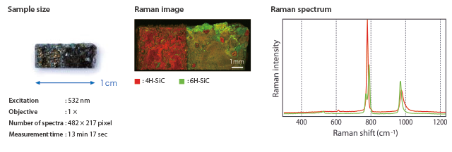
This shows the distribution of polymorphic changes from 4H to 6H of a SiC crystal surface. RAMANview, equipped with wide field-of-view objective lens and fast laser beam scanning, is the best analytical tool for the analysis of the polytypic distribution over a wide range in just 10 minutes.
Crystal quality analysis of diamond
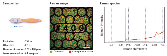
It is an analysis example of diamond file. Diamond has a sharp peak at 1330cm-1, whereas amorphous carbon which is an artificial diamond has a broad peak. It is also applicable to analysis of large-area DLC film.
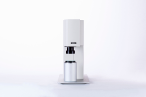
Sophisticated design and usability
The huge workspace is the result of an optimized, reduced to the most constructive and fundamental criteria of design and functionality. Most modern minimalism for highest usability!
Outstanding usability derived from the pursuit of optimized design
Performance and Specification
High quality 532nm laser which gives high performance and a 671nm that is effective to avoid fluorescence. The abundance of lineup and objective lens are correspond to various analysis scene.
Specification
| Wavelength | 532 nm, 671 nm and others |
| Objective lens | 0.5x, 1.0x, 2.0x |
| Illumination | LED reflection |
| Focal length of the spectrograph | 350 mm |
| Grating (selectable with the order) | 600 gr/mm, 1200 gr/mm, 2400 gr/mm |
Performance
| 0.5x objective | 1.0x objective | 2.0x objective | |
| Field-of-view (FOV) | 25.6 x 34mm | 12.8 x 17mm | 6.4 x 8.5mm |
| FOV of Raman imaging | 25 x 25mm | 12.5 x 12.5mm | 6.2 x 6.2mm |
| Spatial resolution | 12 µm | 6 µm | 3 µm |
| Spectrum merasurement range | 100~3200 cm-1 (Option: 50 cm-1~) | ||
| Spectral pixel resolution | 1 cm-1@2400gr/mm, 2 cm-1@1200gr/mm, 4 cm-1@600gr/mm, |
||
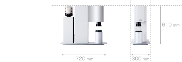
Size and weight
| Size (W × H × D) | 300 × 610 × 720 mm (Baseplate included) |
| Weight | 35 kg (Baseplate included) |