Here, terms are explained in order to get a better understanding of microscope terms that we often see or hear.
Magnification
If a 10 micron sample looks 10 mm, the magnification will be 1000 times.
If it is in the picture, it will be clear because you can hit the ruler. A 10 μm object that is 10 mm in the picture is 1000 times larger.
The magnification during observation is the magnification of the microscope by multiplying the magnification of the objective lens and the magnification of the eyepiece. This magnification is calculated correctly as long as you use an objective and an eyepiece that meets the microscope. (It is designed that way.)
There are microscopes of 160 mm and 210 mm in length depending on the manufacturer, depending on the manufacturer. The eyepiece and the objective lens are made to fit this distance. The magnification is in this design specification, so if you use an objective lens with a different setting, the magnification will be different. Other two lenses are designed so that the best image and performance can be obtained, so it is not recommended to mix and use lenses from various manufacturers for unknown reasons.
Magnification simply means how much you stretch, so it is not a measure of whether the image is sharp or not. Even if you stretch a mountain landscape photograph taken with a digital camera, you will not see the leaves that grow on the mountain, it will be blurry and you won’t know what it is.

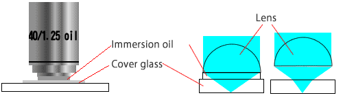
Immersion lens
It is usually an objective lens with a line under the notation of magnification. It is also known as “OIL”.
This lens is filled with an oil with a glass-like refractive index between the lens and the sample to eliminate the effects of air and lens refraction. Therefore, if you do not use oil, performance can not be fully achieved.
With ordinary lenses, the medium that passes light changes in two places: lens (glass) → air → cover glass, and refraction occurs. Oils and immersion oils used in oil immersion lenses have a refractive index that matches that of glass, so refraction does not occur. It is a feeling that the sample is taken into the glass. This leads to an increase in the numerical aperture, which in turn leads to an increase in resolution.
Because of their high performance, oil immersion lenses are also being applied to semiconductor exposure equipment. In addition, even with lenses that do not use light, such as electron microscopes, electron lenses that function similarly are called oil immersion lenses.
Numerical aperture(NA)
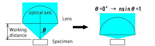
The numerical aperture determines the resolution. Assuming that the angle of the cone that is fully extended to the objective lens from the focal point of the objective lens is θ, the NA and numerical aperture are represented by n sin θ. Here, n is the refractive index of the substance between the objective lens and the sample.
Intuitively, it is how wide you can take in the light from the sample. The lens also shows the numerical aperture, which is about 0.95 for high-performance objectives.
Since the refractive index of air is 1, NA does not exceed 1. Conversely, if n is larger than one, a larger numerical aperture can be obtained. The oil immersion lens has a larger NA by filling an oil or the like with a higher refractive index than air between the objective lens and the sample. The immersion lens has a numerical aperture greater than 1 and in some cases 1.40. The fact that the numerical aperture is 1 or more means that θ is 90 ° and NA = 1. Therefore, intuitively, it is a sense that light reflected in the opposite direction to the objective lens is also collected.
The limit of resolution is wavelength / 2NA in air, and the larger the numerical aperture, the smaller the resolution, which means that you can see the finer things. In addition, the lens with a large NA will necessarily have a short working distance.
Resolution
It is a numerical value that indicates how small things can be seen. The magnification can be increased by increasing it, but if the resolution is not sufficient, it will be blurred when expanded. This is why if you enlarge the image of your camera phone, it will become blurry.
It is also called resolution, but with a microscope, the resolution is the smallest distance between two lines (points) that can be discerned by the device. A resolution of 1 micron means that two lines spaced 1 micron apart look like two lines. The smaller the distance, the higher the resolution and the higher the resolution.
The number of pixels is also a number that represents another feature of resolution. Even with a 5 million-pixel digital camera, if you don’t have the resolution, you can only get blurred photos. However, usually you should have a high resolution lens with a large number of pixels.
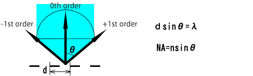
When the slit of the interval d can be determined at the last minute, its resolution is d. The light passing through the slit will be split off as it gets smaller. The light that has passed straight through (0th order) is the light that is always visible, even if the distance is changed. If at least the lowest-order diffracted light (± 1st order) does not enter the lens, it is not clear that it is a slit. The angle θ of diffracted light at this time is d sin θ = λ, where λ is the wavelength. This angle is the same as the definition of the numerical aperture NA, so d = λ / NA. As the wavelength is smaller and the numerical aperture NA is larger, d is smaller, and it can be understood that higher resolution can be obtained.
The above explanation is for the case where the light strikes vertically, but in fact the light strikes also from an angle, so it can be seen that the way of lighting also affects the resolution. Ideally, if the illumination is done with the same lens, twice the resolution is obtained, so the resolution limit is λ / 2NA.
Aberration
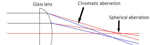
Thin lenses or lenses with long focal lengths do not refract as much and the light is almost in focus, but generally it is out of ideal focus. This shift is called aberration, which causes the image to blur. Aberrations are caused by the fact that light is refracted and the shape of the lens itself.
Chromatic aberration: When sunlight (white light) is passed through a prism and white light passes through glass as the light is split into iridescent colors, the angle of refraction differs depending on the wavelength (color), and the passage path is different. You If you let go through the lens, the color will be faint.
Spherical aberration: The surface of an ordinary lens is spherical, and collimated light entering the periphery and collimated light entering near the center do not focus at the same point. It is a parabolic mirror that the focus is tied to one point. Therefore, for CD and DVD optical pickups, an aspheric lens is used whose curved surface is designed to minimize this distortion. In a microscope, it is a lens with high refractive index, and the curvature is reduced, and the curvature per sheet is reduced by using two lenses to reduce the influence.
Other than this, coma aberration in which the image in the peripheral part is blurred, astigmatism generated in a ray from the outside of the optical axis, curvature of field which can make the image into a plane and curved, and straight lines in the periphery of the screen There are distortions etc. which are bent in the mold.
Aberrations are designed to be small using several lenses, such as changing the material of the glass and combining and canceling lenses of opposite nature. Depending on the type of this correction, microscope objectives include lenses such as “Achromat,” “Apo Chromat,” and “Plan Apo Chromat.”
Achromatic has chromatic aberration removed for only two colors (red and blue), so some colors will be blurred even if it is in focus. However, if you take monochrome photos with monochromatic light, that is enough. Apochromat is a lens that removes chromatic aberration of three colors, and Plan Achromat is a lens that corrects the curvature of field of achromatic. Plan lenses can capture the entire field of view without distortion using photographs or CCDs. And Plan Apochromat is a lens with the above two performances, and of course, it becomes an expensive lens.
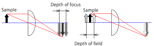
Depth of focus
When focusing on the sample, an image is created on the focal plane. The distance that the image can tolerate (do not blur) when the focal plane is moved is called the depth of focus. Since the depth of focus is inversely proportional to the numerical aperture, we can not say that the resolution is high and the depth of focus is deep. Generally, the higher the magnification, the higher the numerical aperture, the higher the resolution and the shallower the depth of focus. In addition, the depth of focus in photography is only about half of that observed with the naked eye, and it is more difficult to focus in photography than that with the naked eye. The CCD further reduces the depth of focus, but you may focus on it while looking at the monitor, so it may not bother you.
On the other hand, when the sample is in focus, the distance that does not blur even if you move the sample is called the depth of field. Things within the depth of field will be visible at the same time. The depth of field is also inversely proportional to the numerical aperture.
Confocal microscopes have a smaller depth of field than conventional microscopes, but only in focus areas are bright. By using this to shift the sample in the direction of the optical axis (scan in the Z direction) and record only the maximum value of the brightness, you can in principle create an image with an infinite depth of field. Some recent image processing apparatuses select and combine only the images in focus to realize the same function.