Email Magazine
Road to Trial 4(4/28/2022)
This is the fourth in a series of articles challenging amateurs to operate a Raman microscope. Nanophoton’s laser Raman microscope is a device that irradiates a laser beam onto a material to be measured and catches the Raman scattered light coming out of it to read information about the material. The laser has multiple wavelengths, and the user can select which wavelength laser to be installed at the time of installation. Furthermore, it is possible to mount multiple types and switch between them. However, I had no idea why. So this time, I asked an employee to tell me the procedure for switching the laser as a light source and the reason why there are multiple lasers. The key word is “fluorescence. (Takeshi Nemoto, Email newsletter editor-in-chief/freelance writer)
As a layman, it was unclear to me why multiple lasers would be mounted.
When asked by an employee in charge of applications, she said, “When observing various objects or including samples that generate fluorescence, we try to insert multiple light sources (lasers) in advance. If there is fluorescence, the Raman scattering light will be buried by the fluorescence, so they switch the wavelength of the lasers to avoid fluorescence.
Now, let me show you how to operate it while you are actually operating it. Let’s measure a plastic that happens to be nearby.

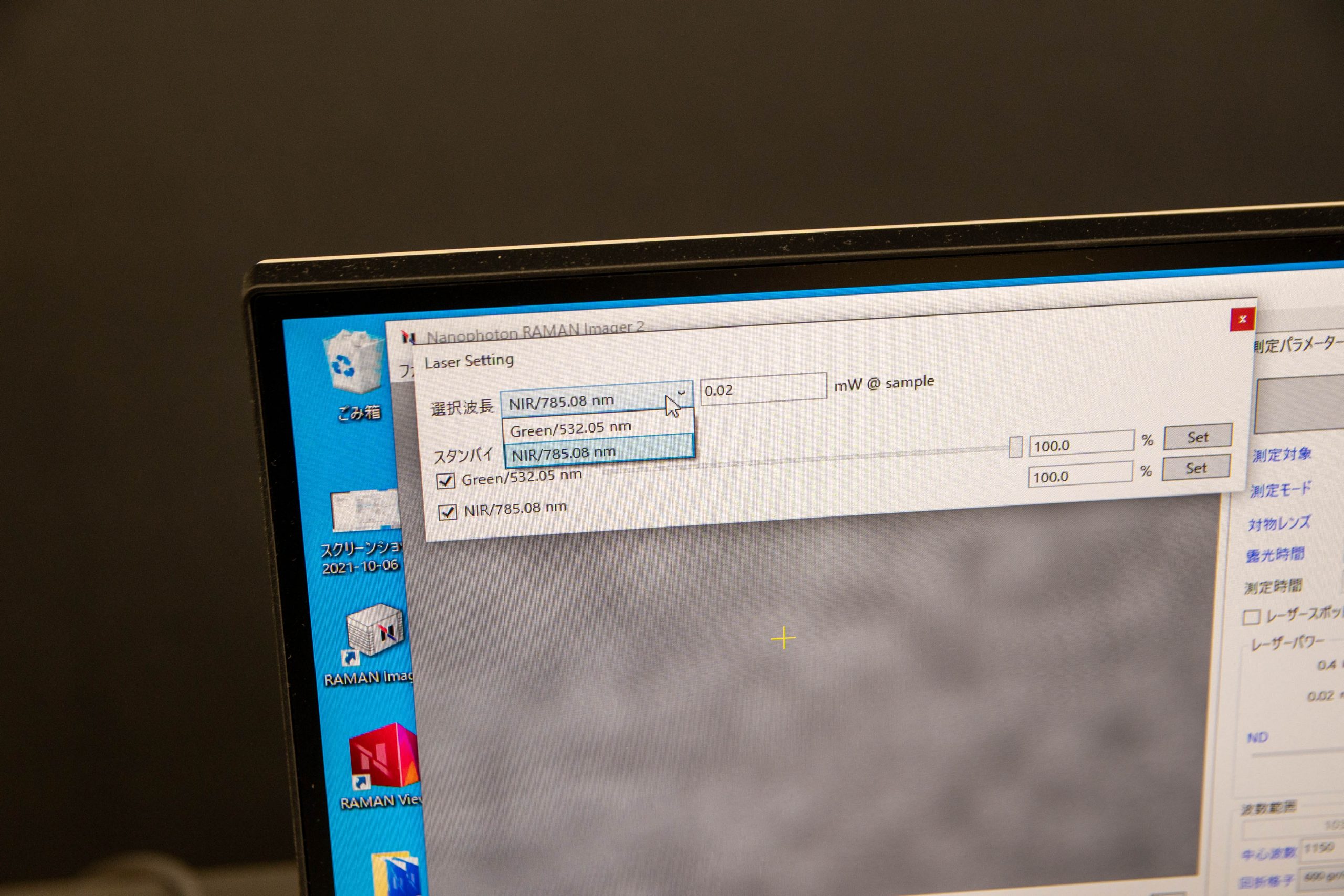
First, let’s take a measurement with a 532nm (green) laser.
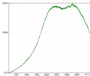
This is a state in which fluorescence has been emitted and Raman scattering light cannot be measured.
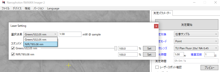
So now we switch to a 785nm (near-infrared) laser and take a measurement. Switching was easy from the software.
After a simple calibration process on the software, I measured the results, which were different from before.
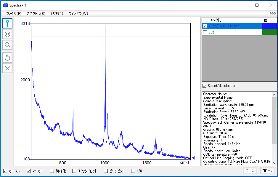
The results of the two laser measurements are compared below. You can see that the Raman scattering light is buried by the fluorescence.

So why can we avoid the effects of fluorescence by changing the laser?
Fluorescence has a fixed wavelength range and appears in the same wavelength range even if the excitation light is changed. On the other hand, the signal obtained from Raman scattering light indicates the energy difference from the excitation light, and if the wavelength of the excitation light source (laser) is changed, the wavelength of Raman scattering light is also shifted. Therefore, if we observe Raman using excitation light in the wavelength range where fluorescence does not appear, we can avoid being disturbed by fluorescence,” says the aforementioned employee.
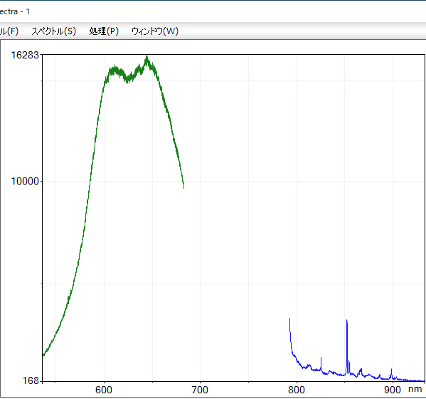
For clarity, let’s look at the spectra observed with two different lasers, with wavelength on the horizontal axis. Green is for a 532nm laser and blue is for a 785nm laser.
When introducing Raman microscopy, it is necessary to select a light source that fits the target sample to be observed. For this reason, the group in charge of applications is trying to propose the best excitation light source by performing demonstration measurements.
Incidentally, when I searched the measured plastic data with the “KnowItAll” software, I immediately realized that it was polystyrene. It is still amazing. I have identified the substance several times with Raman microscopy so far, but the impression has not yet faded.