Email Magazine
Visit to Kyoto University(1/19/2022)
Yasuo Matsumura, a specially appointed professor at Kyoto University, is using a nanophoton Raman microscope for food research. I am very curious to know what kind of research he is doing. I visited Kyoto University’s Uji campus to talk to him. (Email Newsletter editor-in-chief / freelance writer Takeshi Nemoto)
Until last March, Professor Matsumura was a professor at the Graduate School of Agriculture, Kyoto University, where he was involved in food research. He retired there and is now continuing his food research as a specially appointed professor at the university’s Research Institute for Sustainable Humanosphere.
Professor Matsumura: “I am currently affiliated with a laboratory that is leading the research on cellulose nanofibers in Japan. Cellulose nanofibers are made by shredding cellulose, a plant fiber, into nano-sized pieces. I was invited in to see if the application of cellulose nanofibers could be further expanded to the food field.
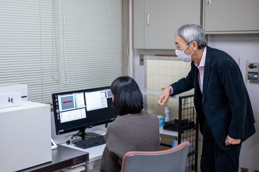
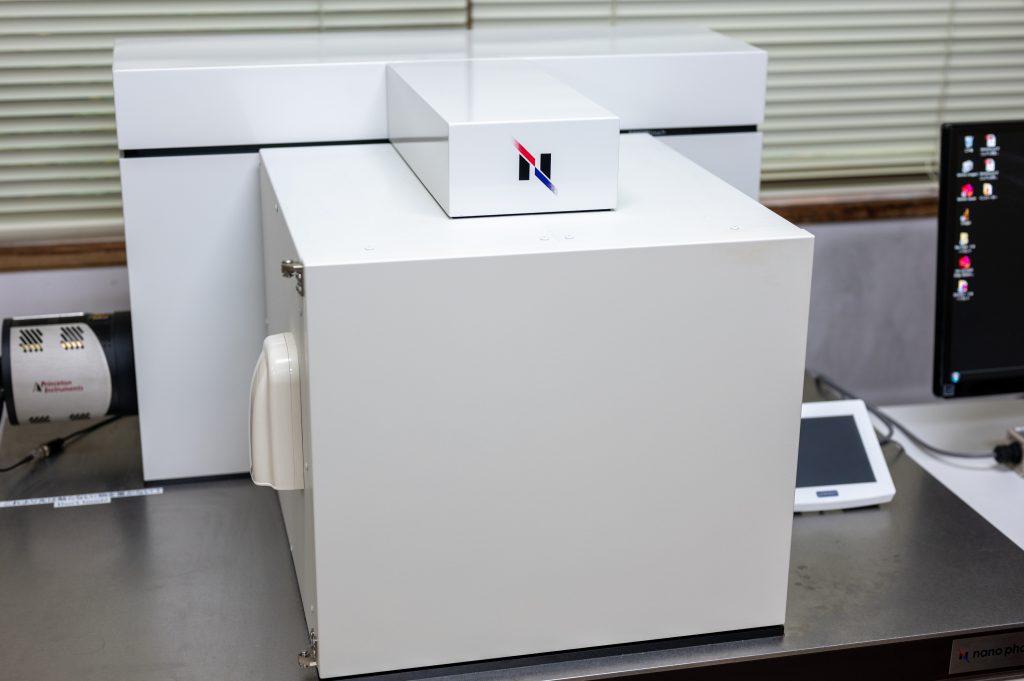
At the Graduate School of Agricultural Science, they were using Nanophoton’s RAMANtouch laser Raman microscope in their research on “dispersed food. By the way, what is dispersed food?
Prof. Matsumura: “Actually, most foods are dispersive foods. One food that is not a dispersant is, for example, a clear juice that contains no solids. There are very few foods that are dissolved in the form of molecules, such as this juice, that can be drunk or eaten. Most foods are in the form of dispersed colloidal particles that are larger than molecules. Milk and mayonnaise are fats and oils (liquid) dispersed in water (liquid), and soups and gels are solids dispersed in a liquid. Bread is a gas dispersed in a solid. These structures affect the stability of food quality and texture.
Both quality stability and texture, such as drinkability and chewability, are important factors for food products. For this reason, Prof. Matsumura often conducts joint research with food manufacturers.
How does Raman microscopy help in these studies?
Prof. Matsumura: “We want to know the relationship between the structure of food and its properties such as quality stability and texture, so we use Raman microscopy as one method. The relationship between structure and properties means, for example, that if fats and oils are well dispersed in a liquid, the quality stability is good, or the drink is pleasant. For example, if fats and oils are well dispersed in a liquid, the quality of the product is stable, or the product is comfortable to drink, or if a solid protein network structure is formed, the product is chewy. Since we are studying how the ingredients interact with each other and how they are distributed, Raman microscopy, which allows us to observe without chemical modification or fixation, is very useful.
As an example, he introduced his research on bread. They are investigating how the types of fats and oils used in the flour dough affect the softness and other characteristics of the bread.
Prof. Matsumura: “When bread is baked, adding oil and fat not only improves the flavor but also makes it soft and puffy. However, there has been little research on how fats and oils are dispersed in the baked product to give it these properties. For this reason, we decided to observe the structure of the dough using a Raman microscope from the dough stage to after it was baked. We were able to distinguish whether the fats were in a solid or liquid state, and found that the properties of the bread changed depending on whether they were solid or liquid.
They plan to publish their research results in a paper in the future, and we look forward to that time.
In addition to this, he has also used a Raman microscope to visualize the way food is cooked as part of an attempt to scientifically understand the characteristics of Japanese food.
Prof. Matsumura: “We decided to study six types of cooking methods (cutting, grinding, boiling or simmering, baking, steaming, and frying). When food is heated during cooking, the state of its components such as protein and water changes. We thought it would be possible to visualize this change in state using Raman microscopy.
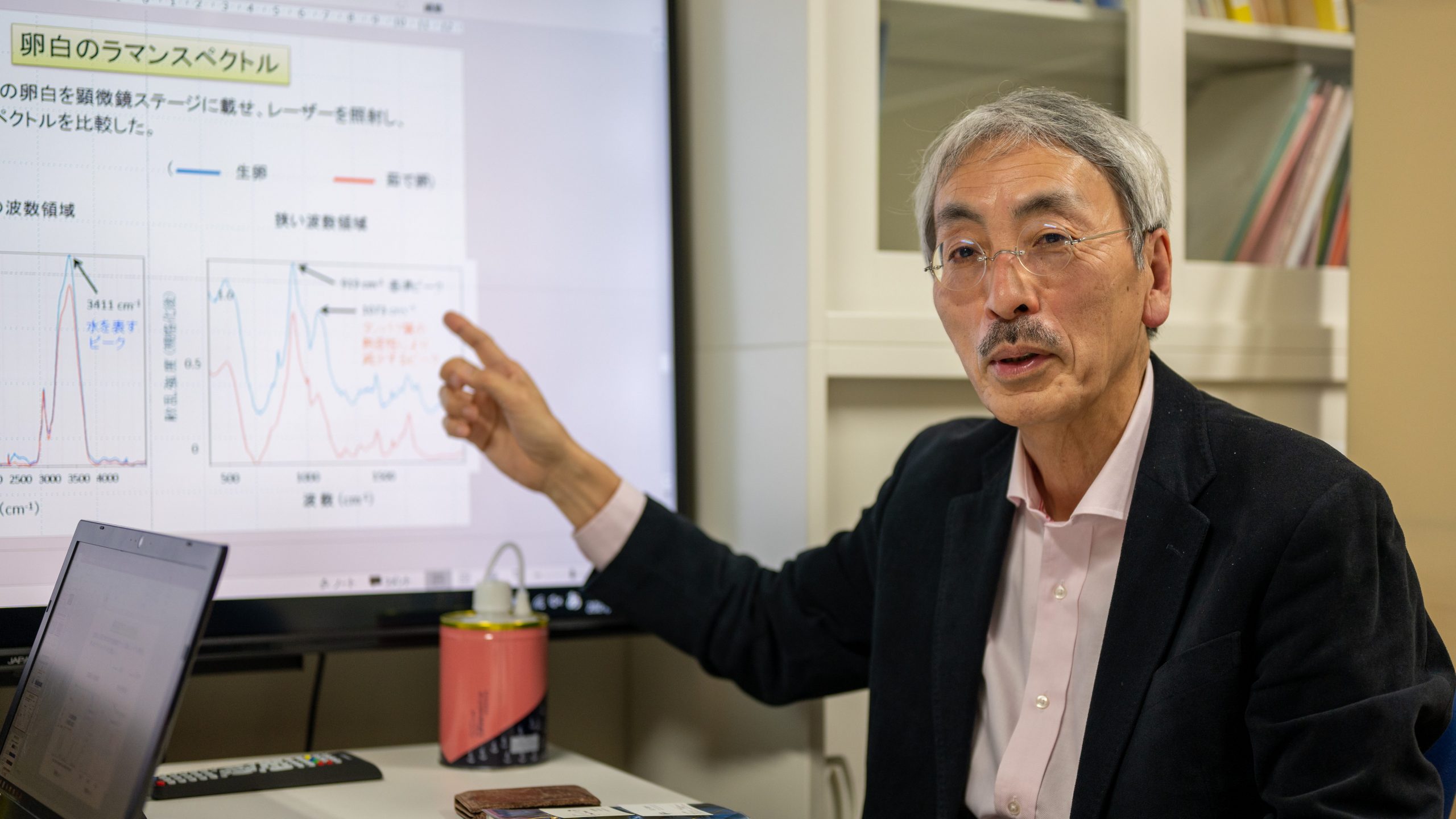
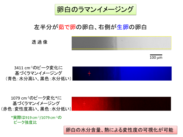
When they observed the egg whites of raw and boiled eggs, they found differences in the peaks of the Raman spectra, as shown in the figure below provided by Prof. Matsumura.
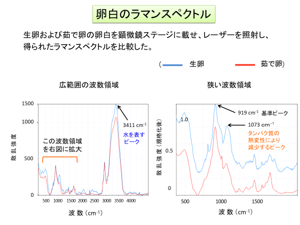
For this reason, Prof. Matsumura and his colleagues performed imaging based on the peak changes at 3411 cm-1 and around 1000 cm-1, and obtained the image shown below. The moisture content and the degree of denaturation due to heat make it possible to visualize how the heat passes through the material.
Prof. Matsumura and his colleagues also compared Raman spectra of three types of sea bream fillets: raw, steamed, and baked, and obtained the following results.

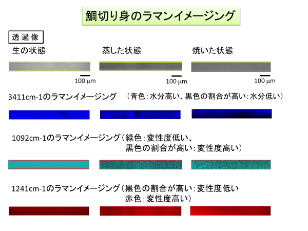
Prof. Matsumura: “By visualizing the cooking process in this way, there is a possibility that we can get closer to the secrets of cooking to find out what the difference is between a master cook and an amateur. Also, for example, when a food processing company develops a food product using fluffy eggs, the visualization by Raman microscope may be useful. Having the Raman microscope as a weapon makes me want to do various kinds of research.
I learned a lot from Prof. Matsumura’s research. I am very much looking forward to his future research.