Email Magazine
Case Studies (4/28/2021)
“Mutant strains of a new coronavirus can be distinguished by Raman microscopy. I was surprised to hear Professor Giuseppe Pezzotti of the Kyoto Institute of Technology say this. When a laser beam is shone on a material, Raman scattered light reflecting differences in structure at the molecular level is emitted. Raman microscopy makes use of this phenomenon, and he said that the differences between the mutant strains of COVID-19 also appear as differences in Raman spectra. This is the first in a series of articles introducing companies and laboratories that have introduced Nanophoton’s Raman microscope. In this issue, we visited the Laboratory of Ceramic Physics at the Graduate School of Science and Technology, Kyoto Institute of Technology (Sakyo-ku, Kyoto City). (e-mail Newsletter editor-in-chief / freelance writer Takeshi Nemoto)
Four doctoral degrees
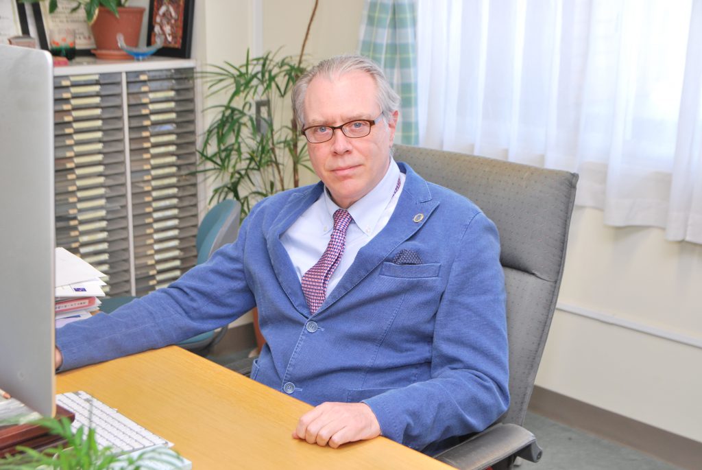
Professor Pezzotti in the lab is from Italy. He is also currently serving as the Vice President of the university. His business card lists him as Doctor of Engineering, Doctor of Medicine, and Doctor of Science.
“In 1987, when I was 27 years old, I came to Osaka University, where I received my doctorate in engineering for my research in materials chemistry. Since 2010, I have been doing research in the medical field, and received my M.D. in orthopedics from Tokyo Medical University, and my M.D. in immunology from Kyoto Prefectural University of Medicine last year. I have a total of four doctorates.
Research in the medical field is conducted using a nanophoton Raman microscope, in collaboration with Professor Osamu Matsuda (Immunology) and Lecturer Tetsuya Adachi (Dental and Oral Sciences) of Kyoto Prefectural University of Medicine.
“Nanophoton’s Raman microscopy is very fast and allows us to see cells, bacteria and viruses alive. The data processing is excellent, and we get beautiful images. Whoever created the software understands the needs of scientists. Fast measurements and clean imaging are amazing advantages in the medical field.”
Raman microscope used in orthopedic and dental research
There are four major research themes that I have pursued using the Raman microscope. Let’s talk about them in order. First, orthopedics. This is the study of cells that make bones.
“We have visualized for the first time at the level of a single cell what kind of metabolic rate the cell uses to make bone. I think this is a great achievement. What we found out is that when we put ceramic in the body, various phenomena occur between the ceramic and the cells. For example, aluminum oxide was generally thought to have no reaction in the body, but in fact it induces cell death through the action of ions. On the contrary, silicon nitride increases the number of cells. In this way, the Raman microscope visualizes and teaches us about phenomena in the microscopic world.”
The next step was in the field of neuroscience. Professor Pezzotti and his colleagues observed Schwann cells, which are responsible for the maintenance of the neuronal network.
“We found that some cells were working and others were slacking off. They look the same under an optical microscope, but under a Raman microscope we could clearly distinguish the differences in cell structure.”
In the field of dentistry, periodontal disease and tooth decay bacteria in the mouth are the targets of research. For example, a substance produced by periodontal disease bacteria has been linked to Alzheimer’s disease and arthritis, and the relationship between this substance and nerve cells, etc. can be examined using Raman microscopy. These are joint researches with Professor Shigetomo Kanamura and Lecturer Toshiro Yamamoto of the Department of Dentistry and Oral Science, Kyoto Prefectural University of Medicine.
Started virus research two and a half years ago.
The research of Prof. Pezzotti and his colleagues is very interesting and I would like to hear more about it, but I will focus on the virus research for this interview. Professor Pezzotti started this research about two and a half years ago with Professor Matsuda’s group at Kyoto Prefectural University of Medicine. This was before COVID-19 infection was known to the world.
“I was interested in the mechanism by which viruses infect cells, and I wanted to visualize it. To do so, I first created a library of Raman spectra of viruses such as influenza with Professor Matsuda.”
If the Raman spectra of various viruses are collected and maintained as a library, it can be used as a database to identify the type of virus. We can measure the Raman spectra of viruses of which we want to know the type, and search the database for viruses with the same characteristics. It is just like identifying an individual with a fingerprint.
COVID-19 is also identified by Raman spectra.
“The Raman spectra of the H1N1 and H3N2 influenza viruses showed unique patterns. For example, there are two types of influenza viruses, H1N1 and H3N2, and the Raman spectra showed a unique pattern for each. Of course, we can determine the difference between the types by analyzing the genes, but what is amazing about Raman is that it does not take much time. Furthermore, it was amazing that the Raman spectrum was able to distinguish the mutant strains of the new coronavirus. No one had done that kind of research yet, and it was unexpected.”

Why is it possible to distinguish between viruses?
“First of all, the ratio of purines (A, G) to pyrimidines (C, U) in the viral genome differs among strains, which is reflected in the intensity of the peaks. There are also differences in the secondary structure of the protein, such as the alpha helix, and the S-S bond. Mutant strains of novel coronaviruses are generally explained only in terms of differences in spike proteins, but in fact, genomes, secondary structures of proteins, and amino acid compositions also differ.”
Professor Pezzotti compares the Raman spectrum to a barcode. All barcodes look the same, but the thickness and spacing of the lines are different, and the information is expressed by these differences. In Raman spectra, various information can be determined by three parameters: the position, strength, and width of the peak.
Determine virus infectivity.
Viruses are very small, and it is difficult to distinguish each one with a Raman microscope. For practical use of virus identification, it is necessary to develop a technology to collect viruses.
On the other hand, it has an advantage over the PCR method currently used to identify viruses: PCR does not tell you the activity of COVID-19, that is, whether or not it is capable of infecting you. Raman microscopy, however, is different.
“We are currently working on the development of materials with antiviral activity against the new coronavirus as part of an AMED (Japan Agency for Medical Research and Development) project. When the antiviral material was exposed to COVID-19, the virus was inactivated and the Raman spectrum changed. With Raman microscopy, we can also determine if the virus is capable of infecting us.”
Professor Pezzotti’s laboratory installed a Raman microscope for Nanophoton’s this year. Until then, they had been using equipment from an external institution at a remote location.
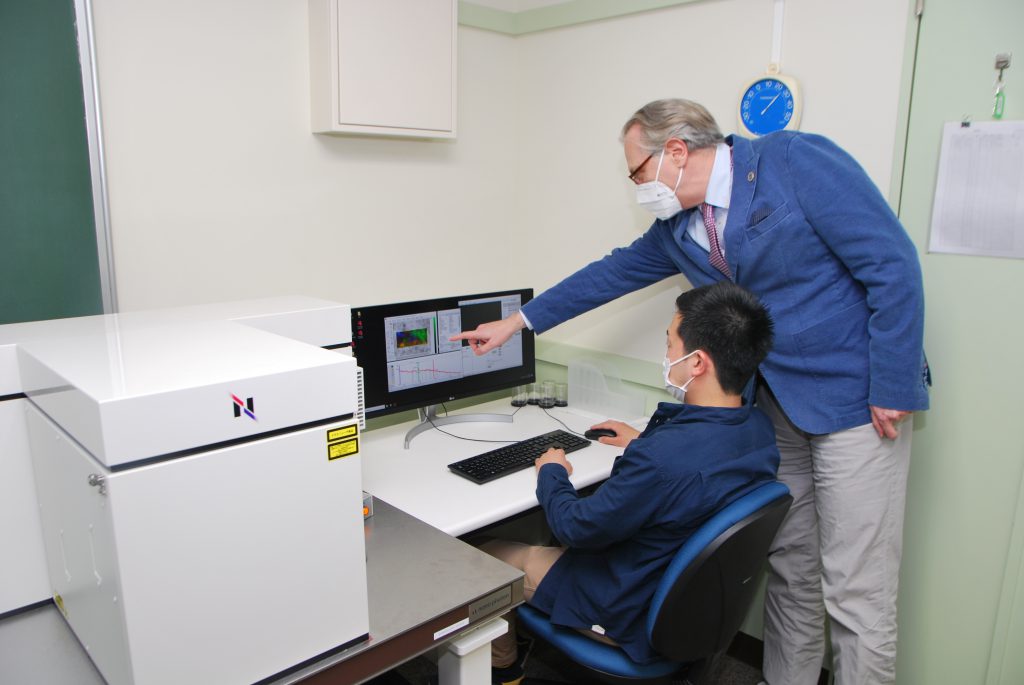
“The Raman microscope has become a mainstay of my research, and borrowing it is no longer enough. With the Raman microscope, cells and viruses whisper to you what they look like. Once you see it, you can’t stop. I want to see it every day, so I bought one.”
“I want to use the Raman microscope to extract all the information.”
I felt his strong enthusiasm for research. With Raman microscopy, we can see the world that no one has ever seen before. I guess that’s what attracts him. “I was impressed by the expression, “it whispers to me.
“I would like to use the Raman microscope to extract all the information. I’m Italian, so when I first saw Chinese characters, I thought, ‘What is this? It’s the same with the Raman spectrum. When you first see it, you think, ‘What is this? That’s my job.”
Lastly, an order to Nanophoton.
“Now that you have invented such a wonderful device, I hope you will invent a portable device. Then we will be able to hear the ‘whispering’ of bacteria and viruses immediately in the medical field.”