Email Magazine
Raman microscope(10/13/2021)
I wish I could use a Raman microscope myself… In this corner, we have asked our employees to measure a variety of everyday objects such as packaging plastics and foods with a Raman microscope. “But I want to measure them by myself. This desire became stronger and stronger, so I asked the company to teach me how to operate it. I asked him to teach me how to operate the device, and he agreed to give me a little training, which I will report on in the next few articles. The first time, I was hesitant to touch the device. It is, after all, a very expensive device. However, it is designed to be easy to use, and it was easier to measure than I expected. (Editor-in-chief of the E-mail newsletter / freelance writer Takeshi Nemoto)
Nearly 30 years ago, I was a graduate student in biophysics, and I had the experience of looking through a microscope at that time. However, my mind and body seem to have forgotten how to use it. So, I am almost a beginner in microscopy. This is the first time for me to operate a Raman microscope.
My instructor is Mariko Adachi, a senior engineer in charge of Sales & Applications. Ms. Adachi said, “My ultimate goal is to be able to measure things around me by myself, to find out what they are made of, and to see how they change under different conditions. Specifically, I want to be able to analyze the composition of the multilayered structure of packaging plastic, or what happens to chocolate when it is boiled at different temperatures. I added, “This is a grand plan, so I want you to proceed little by little”.
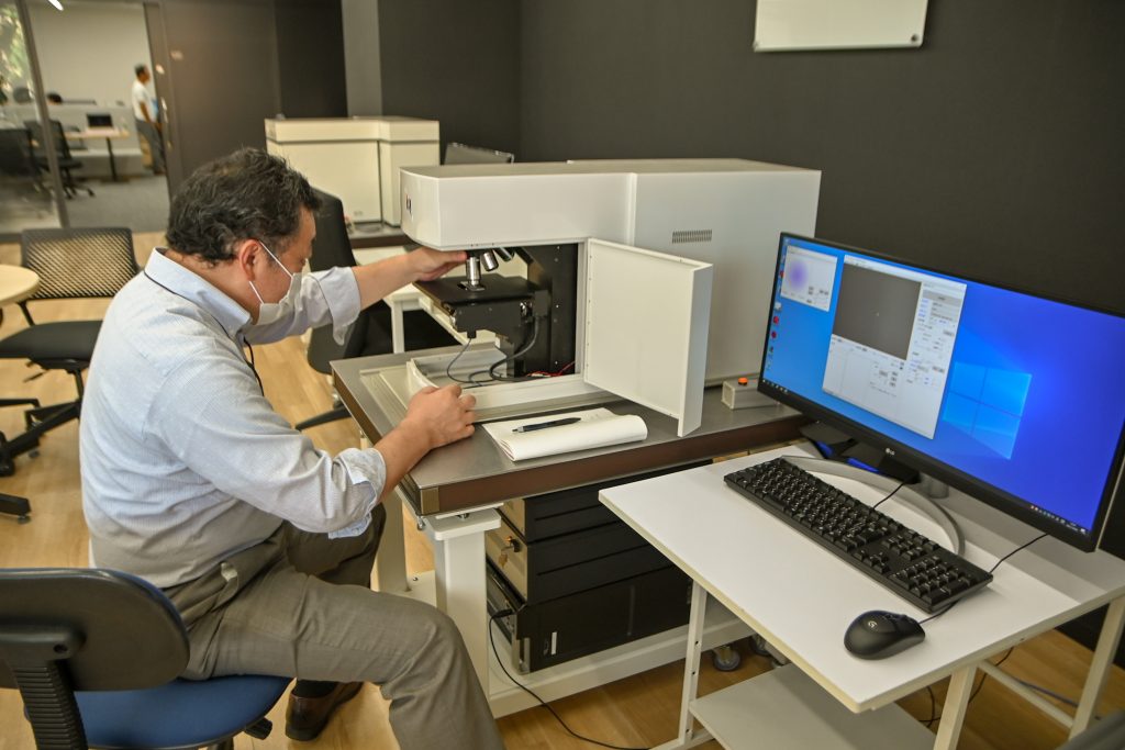
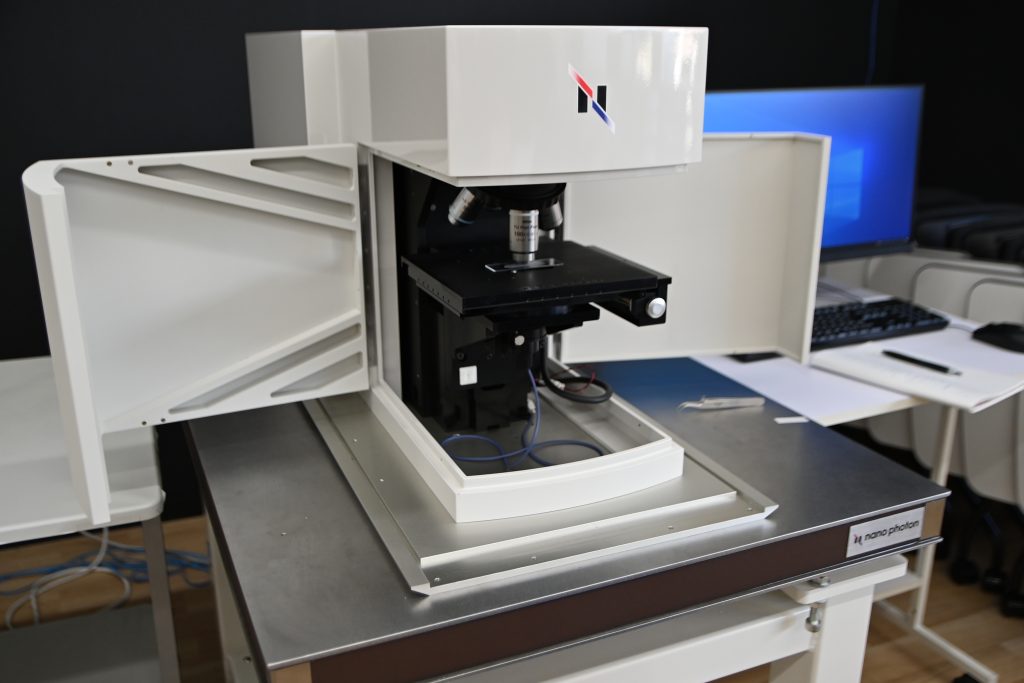
Well, this is the first time. I am going to actually measure samples with the Random Scanning Confocal Raman Microscope RAMANwalk and try to analyze the components. The sample is a clear file prepared by Ms. Adachi.
Place a small cut sample on a glass slide and set it on the stage of the microscope.
The next step is to move the stage up and down to adjust the focus. In the past, when I used a microscope, I first turned the adjustment screw with my right hand to bring the objective lens as close to the preparato as possible, and then looked through the eyepiece to focus. I learned this in junior high school science.
However, Nanophoton’s Raman microscope is different. There is no eyepiece, and it is operated with a mouse wheel while looking at a computer screen.

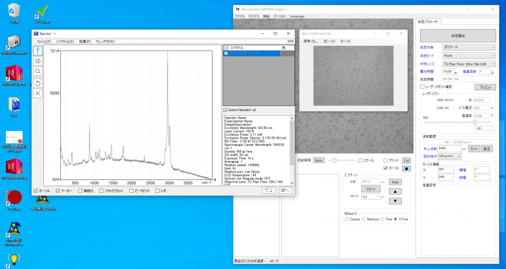
This is what the computer screen looks like.
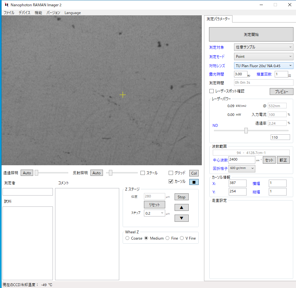
After selecting “Mediem” or “Fine” for “Wheel Z” at the bottom, turn the wheel toward you to lower the stage. If you select “V Fine”, you can move the stage very finely, so use this mode if you want to increase the magnification of the objective lens.

The details of the operation are omitted, but after focusing in this way, it is time for measurement. You need to set parameters such as “laser power”, “exposure time”, “diffraction grating”, “center wavenumber”, etc. Typical settings are registered as “measurement target”. You can start by measuring with the registered settings and then change each parameter as necessary. This time I chose “Polymer H”.
All I had to do was select the point I wanted to measure with the mouse and press the “Start Measurement” button. With almost no waiting, I got a spectrum as shown in the screen below.
After this, I saved the spectra and proceeded to check them against the database of Raman spectra, which I will explain in detail in the next issue of this section.