Email Magazine
ZTrack
Nanophoton’s Raman microscopes have a function called “ZTrack” that can obtain out-of-focus Raman images even of uneven samples. This function measures the unevenness of the surface first, and then automatically changes the focal point of each measurement point using the measured data. However, the employee in charge of applications had a “problem” with this function. When the spatial resolution is high, there will inevitably be dark areas in the Raman image, but this is often intuitively misunderstood. …… What does this mean? We asked for more details. (Takeshi Nemoto, Editor-in-Chief of the Email magazine and science writer)
Before discussing the ZTrack function, please allow me to explain the “confocal optics” used in Nanophoton’s Raman microscopes, as it relates to the development of the ZTrack function.
“RAMANtouch” and “RAMANwalk” Raman microscopes have high spatial resolution in the depth direction and can analyze tomographic and other surfaces with high accuracy. This is also thanks to the confocal optics. I have also used RAMANtouch to analyze a three-layered plastic. The figure below shows the results. The three-layer structure was visualized by color coding in green, red, and blue.
The ability to obtain spectra not only of the sample surface but also of the area below the surface is due to the use of confocal optics, which allows us to focus on the area we want to know about.
The figure below shows how the confocal optics works. A pinhole is placed in front of the detector to cut off excess light from outside the focal plane. This means that Raman scattered light from out-of-focus areas is not detected.
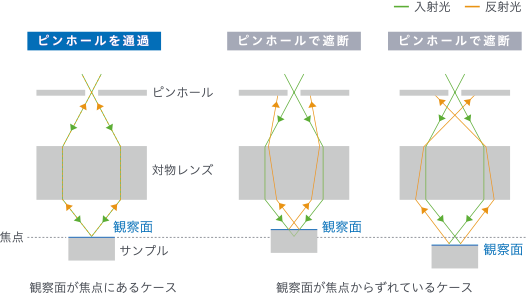
For camera enthusiasts, it may be easier to visualize if we say that it is similar to a case of shallow depth of field. When the area that appears to be in focus (depth of field) is narrow (shallow), the background and foreground are greatly blurred and people and objects can be well emphasized. The confocal optics of the Raman microscope goes even further and excludes out-of-focus areas.
Not detecting out-of-focus areas is very good, but this 3D resolution can backfire if you want to observe the surface of an uneven sample. You can only see the areas that are in focus.
For this reason, before the development of the ZTrack, we were measuring in all three dimensions; we were getting a lot of circular slice images, like a CT scan. But that would have taken a long time. That is why the ZTrack was developed, which first measures the shape of the surface irregularities and then changes the focus according to the shape.
Compare the normal measurement with the ZTrack measurement. They are completely different.

Now, let me preface this, what do you mean by “high spatial resolution inevitably causes dark areas in Raman images, which are often intuitively misunderstood ……”, which is the concern of the employee in charge of applications? I asked him to explain in detail.
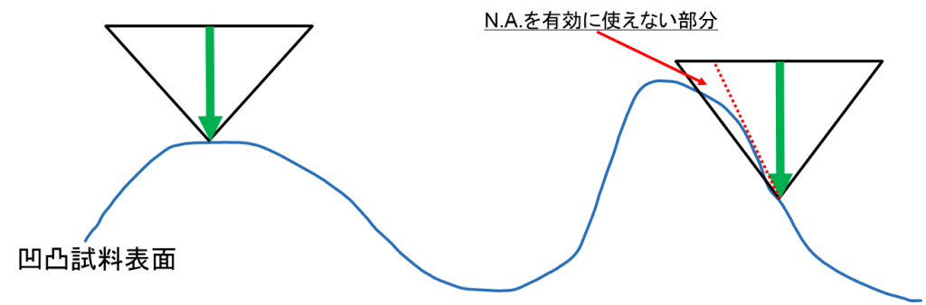
See the figure below. Raman microscope catches Raman scattered light scattered in all directions with an objective lens. What is caught is not all the scattered light, but the light in the area indicated by the ▽ on the right in the figure below.
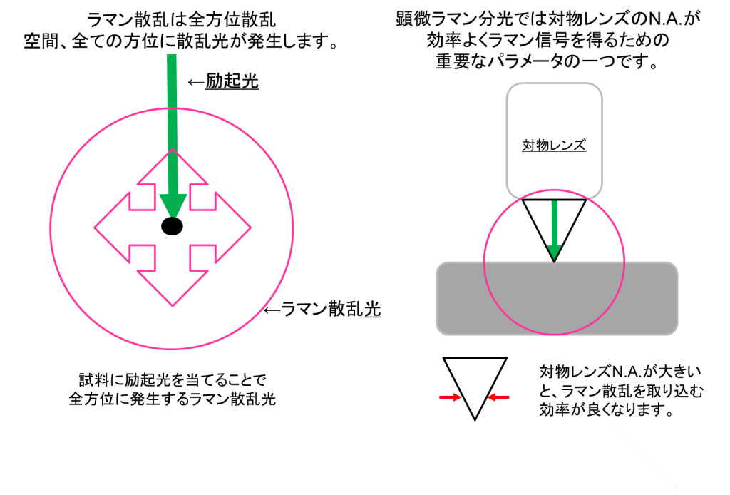
However, on a convex and concave sample surface, as shown in the figure below, this can happen. As shown on the left, it would be good if all of the “▽” could be caught, but on a slope, a part of the surface is in shadow, and the amount of light entering the objective lens is reduced. Therefore, the Raman image of the sloped area becomes darker. Because of this phenomenon, some people say that “Raman image observation of nanophoton’s surface concavo-convexity has dark areas” or “data is missing”.
However, this phenomenon occurs only because of the high spatial resolution of the nanophoton instrument.
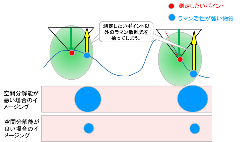
What happens if the spatial resolution is low? As shown in the figure below, Raman scattering light from points other than the one you want to measure is picked up, creating an image as if the sensitivity were high. It is important to note that it is not the Raman scattering light of the point you want to measure.
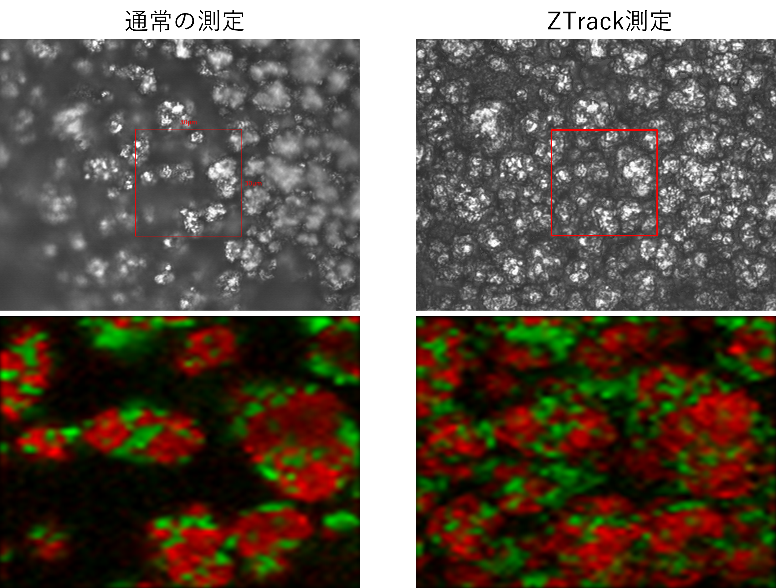
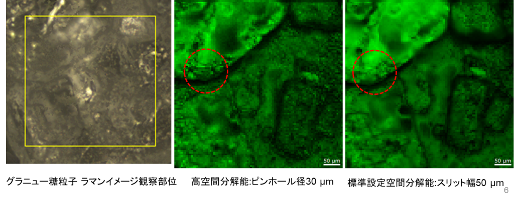
As a test, we measured the same sample with the same instrument with a lower spatial resolution, as shown in the figure below. The sample is granulated sugar particles. The sample with lower spatial resolution (right) has fewer dark areas overall. As shown in the red circle, the bumps on the particle surface can be seen when the spatial resolution is high (center), but they cannot be clearly observed when the spatial resolution is lowered.
Just because an area is dark does not mean that it is not catching Raman scattered light. It is darker because of the relatively low intensity of Raman scattered light. I would appreciate it if you could understand this point,” said the employee in charge of the project. The employee in charge of the project said, “We would appreciate it if people understood this point.